Introduction
Studies of sensory perception and motor control is an exciting area of investigation in neuroscience and physiology. Two key players involved are mechanoreceptors and proprioceptors; both play key roles in sensing physical stimuli but may differ in terms of location, function and types of information they provide to the central nervous system.
Mechanoreceptors are sensory receptors found throughout our bodies, most notably skin and subcutaneous tissues. Their purpose is to detect various mechanical stimuli like pressure, touch, vibration and texture – when activated they convert these mechanical signals into electrical impulses that travel to our brain for interpretation, helping us perceive and respond to tactile sensations in the environment.
Proprioceptors, on the other hand, are sensory receptors found within muscles, tendons, joints, and inner ear structures that serve to give us information about body parts’ positions, movements, tension levels and tension changes. Proprioceptors play an integral part in body awareness by providing us with accurate sensory perception of where limbs, joints and overall body positions lie within space.
Reducing confusion about sensory integration within the central nervous system by distinguishing mechanoreceptors and proprioceptors is essential to understanding how different sensory data is processed and integrated within our bodies.
By studying their characteristics, functions, and neural pathways associated with each type of receptor we gain valuable insight into how our bodies perceive physical stimuli, maintain balance and coordination, or execute precise motor movements.
We will investigate the definitions, characteristics, functions and implications of mechanoreceptors and proprioceptors – with particular attention paid to their similarities and differences – while exploring their applications to sensory perception, motor control, balance as well as clinical applications such as rehabilitation and sports performance.
Eventually we will recognize their significance for furthering knowledge of human sensory and motor systems.
Definition of mechanoreceptors
Mechanoreceptors are sensory receptors that detect mechanical stimuli, including pressure, touch, vibration and stretching. They can be found Throughout the body’s skin, muscles, Tendons and other tissues and convert Mechanical stimulation to Electrical signals which are then Processed and Interpreted by the central Nervous system.
These receptors are designed to detect and relay information about physical forces acting on our bodies, enabling us to perceive and respond to various tactile sensations. Different mechanoreceptor types are responsible for specific aspects of touch perception such as pressure, texture and vibration – each has unique structural and functional features which enable it to detect specific types of mechanical stimuli and transduce this data back.
Mechanoreceptors play an essential role in our everyday interactions with the environment, helping us navigate objects, feel sensations, maintain balance and posture and stay balanced and upright. Deliberating on mechanoreceptors’ role and functioning is integral to fields such as neuroscience, physiology, rehabilitation and sensory perception research.
Definition of proprioceptors
Proprioceptors are sensory receptors that provide information about the position, movement and tension of our body parts. Located mostly Within muscles, tendons, joints and inner ear Structures, Proprioceptors play a central role in helping us Perceive and control the spatial Orientation and motion of our body.
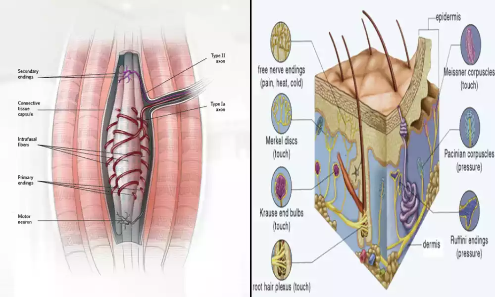
Proprioceptors serve various functions and locations within the body. Muscle spindles, for instance, detect changes in muscle length to enhance our sense of stretching and contraction; Golgi tendon organs offer feedback regarding muscle tension as well as help protect against excessive force on muscles.
Joint receptors are proprioceptors found within the capsules and ligaments surrounding joints that sense changes in joint position or movement, providing us with our sense of its angle or direction. Joint receptors play an especially crucial role in coordinating movement while maintaining joint stability.
Proprioceptors send information directly to the central nervous system, where it combines with other sensory input to form an accurate perception of body position and movement. This proprioceptive feedback plays a vital role in motor control allowing us to perform precise movements while maintaining balance in our surroundings and performing coordinated movements in harmony with one another.
Proprioception plays an integral part in multiple fields, such as sports performance, rehabilitation and movement sciences. Understanding proprioceptors’ function within the body helps in diagnosing and treating movement disorders as well as developing effective rehabilitation programs and improving athletic performance.
Comparison Table of Mechanoreceptors and Proprioceptors
Sure! Here’s a comparison table highlighting the key differences between mechanoreceptors and proprioceptors:
| Aspect | Mechanoreceptors | Proprioceptors |
|---|---|---|
| Location | Primarily in the skin and underlying tissues | Within muscles, tendons, joints, and inner ear |
| Sensory Perception | Touch, pressure, vibration, texture | Body position, movement, muscle tension |
| Types | Pacinian corpuscles, Meissner’s corpuscles, Merkel’s discs, Ruffini endings | Muscle spindles, Golgi tendon organs, joint receptors |
| Function | Detecting mechanical stimuli | Providing information on body position, movement, and tension |
| Neural Pathways | Signals transmitted via peripheral nerves | Signals transmitted via peripheral nerves |
| Interaction and Integration | Contribute to tactile sensations, spatial perception, and texture discrimination | Essential for motor control, coordination, and proprioceptive awareness |
| Importance | Perceiving touch and texture, interpreting external stimuli | Maintaining balance, executing precise movements, and body awareness |
| Clinical Applications | Rehabilitation, sensory research, tactile perception studies | Sports performance, movement disorders, physical therapy |
This table provides a concise overview of the main differences between mechanoreceptors and proprioceptors, including their locations, sensory perceptions, types, functions, neural pathways, and clinical applications.
It demonstrates how these receptors serve distinct roles in our sensory perception, motor control, and overall body awareness.
Importance of understanding the difference between Mechanoreceptors and Proprioceptors
Understanding the difference between mechanoreceptors and proprioceptors is of vital importance for various reasons:
Sensory Perception: Mechanoreceptors and proprioceptors play key roles in sensory perception. Mechanoreceptors detect tactile sensations such as touch, pressure and texture allowing us to interact and explore our environment while proprioceptors provide information about body position, movement and muscle tension which enables us to sense spatial orientation as well as execute precise motor movements. Understanding their distinct contributions aids our comprehension of sensory perception’s complexities.
Motor Control and Coordination: Proprioceptors play a vital role in motor control and coordination, providing constant feedback about muscle positions and tension to enable precise control and adjustment of movements.
By understanding proprioception, we gain insights into how our brain integrates this sensory data with other sensory signals to produce coordinated movements while keeping balance. This knowledge is key for sports performance enhancement, physical therapy and rehabilitation – fields where improving motor control and coordination is often at the core.
Clinical Applications: Understanding the difference between mechanoreceptors and proprioceptors can have far-reaching implications in various clinical settings. For instance, rehabilitation involves understanding proprioception to better assess and treat movement disorders, balance issues, proprioceptive deficits, as well as develop targeted interventions to restore proprioceptive function and enhance motor recovery.
Meanwhile in sports performance mechanoreceptors play a vital role in touch perception which inform training techniques and equipment design that optimize performance while decreasing injury risks.
Research and Innovation: Expanding our understanding of mechanoreceptors and proprioceptors can lead to new discoveries and innovations. Examining these receptors reveals intricate neural pathways involved in sensory perception and motor control; provides insights into the brain’s processes for processing sensory information; opens doors for advancements in fields like robotics, haptic technologies, virtual reality and neurorehabilitation as well as improved human-machine interfaces and sensory feedback systems.
Understanding the difference between mechanoreceptors and proprioceptors is critical to comprehending sensory perception, motor control, body awareness, rehabilitation, sports performance and sensory research – as well as supporting scientific advancement and innovation by unlocking the complex interactions of human sensory and motor systems.
Location and distribution in the body
Mechanoreceptor Location and Distribution:
Mechanoreceptors can be found throughout the body in various tissues and organs. Here are a few key locations and distributions of mechanoreceptors.
Skin: Mechanoreceptors in the skin detect touch, pressure, vibration and texture. They are more concentrated in areas with high levels of sensitivity such as fingertips, lips and palms; however they are present elsewhere but in lesser concentrations.
Subcutaneous Tissues: Mechanoreceptors can also be found in the subcutaneous tissues beneath the skin, where they help the brain register pressure and deep touch sensations. These receptors contribute to pressure perception as well as deep touch sensations.
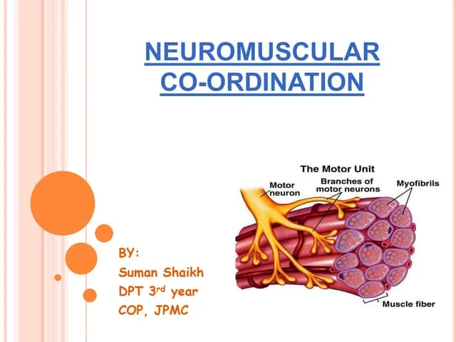
Mechanoreceptors in Skeletal Muscles: Mechanoreceptors found within skeletal muscles play an integral part of muscle proprioception by providing information regarding length, tension, and contraction that allows us to detect and control muscle movements.
Tendons: Some mechanoreceptors are located within tendons that connect muscles to bones, providing feedback about muscle loading and helping prevent excessive strain. These receptors serve to detect tension or force exerted on tendons as a whole and respond by sensing tension or force exerted on them – providing feedback about muscle loading or overload and providing protection from unnecessary strain.
Mechanoreceptors are present in joint capsules and ligaments to provide information about joint position, movement, and mechanical forces acting upon it. Mechanoreceptors help promote proprioceptive awareness and joint stability.
Inner Ear: Inside the inner ear are mechanoreceptors known as hair cells which detect soundwaves and head movement; these mechanoreceptors play an integral part in auditory perception and balance. Mechanoreceptors are located throughout different body regions to meet specific sensory needs and functions associated with those areas.
Distribution of Proprioceptors:
Proprioceptors can be found throughout muscles, tendons, joints and inner ear structures. Here is an outline of their location:
Muscles: Proprioceptors known as muscle spindles can be found within skeletal muscles and are distributed along muscle fibers in their belly area, similar to proprioceptors found on our faces and wrists. Their exact distribution varies according to each muscle’s function and complexity.
Tendons: Proprioceptors known as Golgi tendon organs can be found within tendons where they connect to muscles. Their location near this interface gives information about tension and force during muscle contractions.
Proprioceptors Are Present Within Joint Caps and Ligaments: Joint proprioceptors can be found within joint capsules, ligaments and other structures surrounding joints to detect any changes in joint position, movement and mechanical stress – providing feedback about joint angle or movement.
Inner Ear: Vestibular receptors in our inner ears, known as proprioceptors, detect head movements and changes in body position relative to gravity – contributing significantly to our sense of balance and spatial orientation.
Proprioceptors are distributed throughout the body to provide accurate feedback for maintaining posture, coordinating movements, and perceiving body position. Mechanoreceptors and proprioceptors are present throughout our bodies, helping us perceive mechanical stimuli while keeping body awareness intact.
Neural pathways involved in transmitting mechanoreceptor signals
Mechanoreceptor signals travel along an intricate network of neural pathways. Here are the primary ones involved in transmitting mechanoreceptor signals:
Peripheral Nerves: Mechanoreceptor signals are initially transmitted through peripheral nerves. Peripheral nerves consist of sensory nerve fibers that connect mechanoreceptors located throughout the body to spinal cord and brain; these sensory fibers carry electrical signals produced by mechanoreceptors to the central nervous system.
Dorsal Root Ganglia: Sensory information from mechanoreceptors is relayed through dorsal root ganglia, clusters of sensory cell bodies located along the spinal cord that serve as gateways for sensory information to enter.
Spinal Cord: Mechanoreceptor signals travel from the dorsal root ganglia through sensory nerve fibers to reach the spinal cord, where they are processed and integrated before being transmitted to higher brain centers. Thus, this part of the central nervous system plays a critical role in filtering and modulating sensory information before reaching higher brain centers.
Ascending Pathways: Once in the spinal cord, mechanoreceptor signals are transmitted to the brain via ascending pathways. There are various ascending pathways involved with relaying mechanoreceptor signals to the brain – these include dorsal column-medial lemniscal pathway and spinothalamic pathway.
Dorsal Column-Medial Lemniscal Pathway: This pathway transmits fine touch, vibration and proprioceptive information from mechanoreceptors located in skin, muscles and joints to mechanoreceptors located elsewhere on the brainstem in medulla before ascending through dorsal columns to thalamus for processing by primary somatosensory cortex.
Spinothalamic Pathway: This pathway relays information regarding touch, pressure, temperature and pain from mechanoreceptors. Signals rise up the spinal cord’s anterior and lateral columns before crossing over shortly thereafter into its opposite side shortly before ascending further through to reach the thalamus where they’re relayed onto specific regions of the somatosensory cortex.
Brain Processing: Once mechanoreceptor signals reach the brain, they are processed and interpreted by different parts of the somatosensory cortex. While primary somatosensory cortex serves as the central point for receiving and mapping sensory data, other areas contribute to further analysis, integration, and perception of tactile sensations.
Neural pathways responsible for transmitting mechanoreceptor signals enable humans to perceive and interpret touch, pressure, vibration and other sensory experiences. Signals travel from mechanoreceptors through peripheral nerves, spinal cord and ascending pathways into higher brain centers where they are processed into tangible sensory experiences that lead to our tactile senses.
Providing information about body position and movement
Proprioceptors play an essential role in providing information about body position and movement. Their contributions contribute to our sense of proprioception – our ability to sense body parts’ locations, orientation, and movement. Here’s how proprioceptors provide this data:
Muscle Spindles: Muscle spindles are proprioceptors found within skeletal muscles that detect changes in muscle length and provide essential proprioceptive awareness. When muscles stretch or contract, muscle spindles detect these length changes and communicate that information to our central nervous systems to create an awareness of our own muscles’ position and movement allowing us to gain an awareness of limb placement and movement.
Golgi Tendon Organs: Golgi tendon organs are proprioceptors located within tendons that connect muscles to bones. They detect changes in muscle tension and force by sensing how tension exerts on a tendon during muscle contraction, producing signals to inform the central nervous system about increased tension on a tendon during contractions;
when tension rises further they send feedback signals back into the central nervous system about force exerted during muscular activity – this feedback loop helps control muscle contraction without leading to potential injuries such as overexertion which could cause muscle fatigue and injury.
Joint Receptors: Proprioceptors can be found within capsules and ligaments surrounding joints, providing information about joint position, movement, mechanical stress and awareness of angle changes in joint movement. By sensing mechanical forces acting upon them they help maintain joint stability while coordinating movements.
Proprioceptor information is processed within the central nervous system and processed by areas like the cerebellum and somatosensory cortex of the brain, where it provides coherent perceptions of body position, movement, and spatial orientation.
Proprioceptive feedback allows precise motor control, coordination of movements, posture modifications based on sensory information received via proprioceptors as well as ability to adapt posture or limb positions according to proprioceptor input.
Understanding proprioceptor information is vital for maintaining balance, performing precise movements, and having body awareness. Proprioception helps us perform daily activities such as sports and tasks that require coordinated and controlled movements of our body as well as any necessary rehabilitation or sports performance training sessions that might need coordinated movements of our body parts.
Furthermore, deficits or impairments of proprioception may interfere with motor function leading to movement disorders which highlights its significance in fields like rehabilitation, sports performance analysis and movement sciences.
Regulating muscle contraction and coordination
Proprioceptors play an essential role in regulating muscle contraction and coordination. Their feedback to the central nervous system allows precise control and coordination of muscular activity.
Here’s how proprioceptors contribute:
Muscle Spindles: Muscle spindles, proprioceptors found within skeletal muscles, play an invaluable role in regulating muscle contraction. By monitoring changes to muscle length and rate of change, muscle spindles play an integral part in maintaining tone and stability by alerting spinal cord of any stretch or sudden lengthening in order to activate stretch reflex.
When this occurs, muscle spindles send signals back through spinal cord which activate reflex known as stretch reflex and contract in response causing rapid adjustments as needed as well as overstretching from occurring or rapid lengthening by sending signals back through cord to spinal cord.
which activate stretch reflex causing it to contract in response resulting in improved tone/stabilty as well as rapid length changes/length changes/any adjustments made. This reflex also serves preventing overstretching and rapid length changes while providing quick adjustments due to changes in length or changes due to changes in length changes through quick adjustments on changes occurring.
Golgi Tendon Organs: These proprioceptors located within tendons serve to detect changes in muscle tension and force. When tension in a muscle increases during forceful contraction, for instance during an aggressive set, Golgi tendon organs become activated, sending inhibitory signals back to the spinal cord and initiating what’s known as the tendon reflex.
leading to relaxation of muscles and reduction of force to protect them against potential injury or damage. This mechanism safeguards against damage or injury to muscle fibers from too much force without damaging them further!
Coordination of Muscle Groups: Proprioception helps coordinate muscle groups during complex movements by providing continuous feedback about muscle length, tension and position. Proprioceptors support activation timing across different muscles involved in an action to ensure smooth and efficient movements patterns while avoiding unintended muscle contractions or imbalances.
Integrating proprioceptive feedback with other sensory data, such as visual input, is integral for fine motor control and coordination. The central nervous system utilizes proprioceptor data to adjust contraction levels, modulate tone levels and coordinate muscle activity across body parts – essential elements in performing skilled movements, maintaining balance or performing precise motor tasks.
Disruptions to proprioception due to injury or neurological conditions can lead to coordination deficits, muscle imbalances and difficulty with motor control. Therefore, understanding the role proprioceptors play in regulating muscle contraction and coordination is key for rehabilitation, sports performance and motor learning – interventions can be tailored specifically to boost proprioceptive feedback for improved movement quality.
Neural pathways involved in transmitting proprioceptor signals
Transmission of proprioceptor signals relies on certain neural pathways that connect proprioceptors to the central nervous system.
Here are some key pathways involved in transmitting proprioceptor signals:
Dorsal Root Ganglia and Peripheral Nerves: Proprioceptive signals begin their journey in proprioceptors located within muscles, tendons, and joints. Once processed by sensory nerve fibers carrying information from these receptors, these peripheral nerves combine together into peripheral nerve bundles which then transmit those signals onward to dorsal root ganglia clusters near the spinal cord for transmission to dorsal root ganglia (clusters of sensory cell bodies located close by).
Dorsal Columns: Proprioceptive signals travel up the spinal cord via two major tracts in the dorsal columns that carry proprioceptive information: the anterior column (APC) and dorsal column (DC).
Fasciculus Gracilis: This tract transmits proprioceptive signals from the lower body, such as legs and trunk.
Fasciculus Cuneatus: This nerve tract transmits proprioceptive information from various parts of the upper body, including arms, neck, and shoulders.Dorsal columns exhibit signals which remain on one side of the body; thus enabling accurate readings.
Medulla Oblongata: When proprioceptive signals reach the medulla oblongata, they undergo decussation – in which signals cross over to opposite sides of the brainstem and ascend towards the brain via decussation.
Thalamus: Proprioceptive signals travel next through the thalamus to reach their final destination – specifically through ventral posterolateral nucleus (VPL) for lower body signals and ventral posteromedial nucleus (VPM) for upper body signals. Thalimus acts as a gateway by filtering and modulating this proprioceptive information before it reaches cerebral cortex.
Proprioceptive Signals Are Projected From the Thalamus To the Primary Somatosensory Cortex: Proprioceptive signals generated in the thalamus are sent directly to this region of cerebral cortex responsible for processing and interpreting sensory data, including proprioception. The Primary Somatosensory Cortex also creates a spatial representation of body awareness as well as perceive proprioceptive sensations.
Motor Cortex and Motor Pathways: Proprioceptive signals interact with the motor cortex, which is responsible for planning and executing voluntary movements. Proprioceptive feedback allows this area to adjust and coordinate muscle activity for precise motor control and movement coordination.
Proprioceptor neural pathways provide a means of communicating information regarding body position, movement and tension to the central nervous system. Through integration of proprioceptive feedback with other sensory data sources, they allow brains to form coherent maps of body space orientation while also managing muscle contraction and coordinating movements effectively.