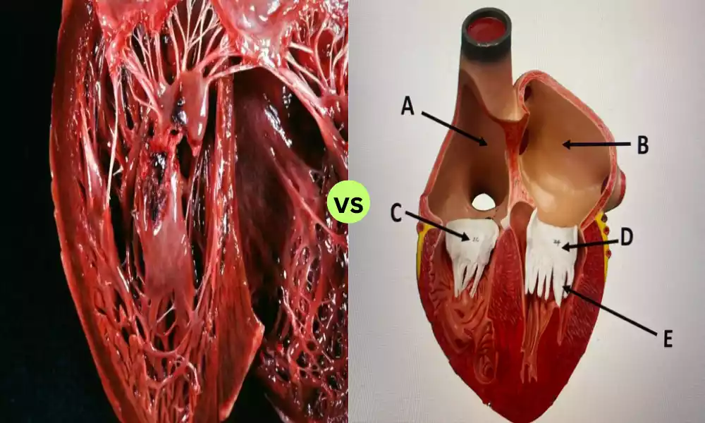
Within the heart’s complex anatomy lies two vital muscle types – Papillary and Pectinate muscles – both essential for cardiac health but located in different regions and serving distinct purposes.
We will examine their differences to reveal their structures, functions, and clinical relevance; understanding which is which provides valuable insight into understanding what drives human existence and longevity.
Definition of Papillary Muscles
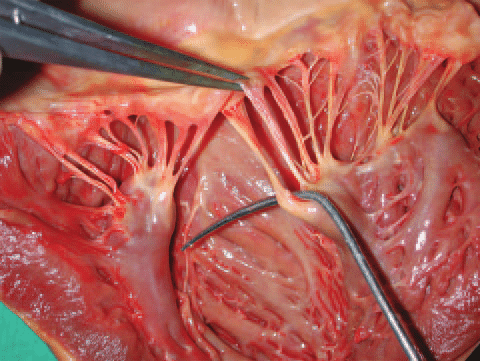
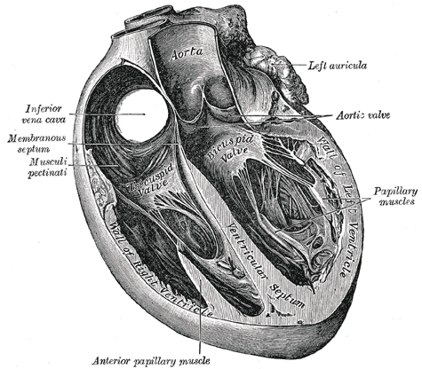
Papillary muscles are tiny muscles that look like fingers and are located in the ventricles of the heart, with a predominant location located in the left ventricle. These muscles play an important function within the cardiac system by connecting to the valves of the atrioventricular (mitral as well as tricuspid valves) via chordae tendineae which are fibrous cords.
Their primary purpose is to stop the prolapse or inversion of these valves into the atria when there is a contraction of the ventricular.
The muscles of the papillary contract before the ventricles contract, tensioning the chordae tendineae and securing the valve leaflets into their place. This process ensures a unidirectional circulation of blood through the heart and is vital to ensure proper cardiac function.
Definition of Pectinate Muscles
Pectinate muscles, also known as muscle pectinati are muscular structures that reside within the heart’s atria and are primarily located inside the right atrium. They have a distinct look that resembles a ridge or comb with muscle bundles that form the interior walls of the atrium, especially in the auricles, which are tiny extensions of the atria like earlobes.
The role of the pectinate muscle is to aid in atrial contraction, especially during the last stage of diastole in the ventricular. This is a way of filling the ventricles of blood, contributing to the effectiveness of the pumping.
Pectinate muscles are involved in “the “atrial kick,” which boosts the volume of blood that flows into the ventricles in each cycle of cardiac activity, which increases the output of the heart.
Comparison Table of Papillary and Pectinate Muscles
Here is a comparison table highlighting the key differences between Papillary and Pectinate muscles:
| Characteristic | Papillary Muscles | Pectinate Muscles |
|---|---|---|
| Location | Found in the ventricles of the heart, primarily in the left ventricle. | Located in the atria of the heart, primarily in the right atrium, especially in the auricles. |
| Structure and Appearance | Finger-like projections that resemble small columns of muscle. | Comb-like or ridge-like muscular bundles, give a serrated or ridged appearance. |
| Attachment | Attach to the atrioventricular valves (mitral and tricuspid valves) via chordae tendineae. | Attach to the inner walls of the atria and the auricles. |
| Primary Function | Prevent prolapse of the atrioventricular valves by stabilizing the valve leaflets during ventricular contraction. | Facilitate atrial contraction, contributing to the “atrial kick” and assisting in ventricular filling. |
| Role in Cardiac Physiology | Ensures unidirectional blood flow through the heart and supports valve function. | Increases efficiency by enhancing ventricular filling and optimizing cardiac output. |
| Clinical Significance | Associated with conditions such as mitral valve prolapse and papillary muscle dysfunction. | Relevant in conditions like atrial fibrillation and atrial flutter. |
This comparison table summarizes the key distinctions between Papillary and Pectinate muscles, highlighting their respective locations, structures, functions, and clinical implications within the cardiac system.
The attachment points to the atrioventricular valves
The points of attachment of those Papillary muscles and Pectinate muscle to the valves for the atrioventricular in the heart can be described in the following order:
Papillary Muscles:
- Papillary muscles are connected to the atrioventricular (AV) valves. These comprise the mitral (bicuspid) valve located on the right side of the heart as well as the tricuspid valve located on the left side.
- Particularly, the papillary muscles connect with the leaflets of valves (also called flaps or cusps) of the valves by fibrous cords, also known by the name chordae tendineae.
- The chordae tendineae stretch from the muscles of the papillary to the edges of the free or border of valve leaflets.
- The tension and attachment provided by the papillary muscles as well as chordae tendineae stop the prolapse or inversion of AV valve leaflets to the Atria during ventricular contraction, making sure that blood flow is one-way from the atria into the ventricles.
Pectinate Muscles:
- Pectinate muscles lack an immediate attachment point to the valves of the atrioventricular as do papillary muscles.
- The pectinate muscle is found in the atria most notably located in the right atrium as well as its auricles (small extensions of the ear).
- Their role within the atria is to assist in atrial contraction. This contributes to the filling of the ventricular cavity.
- Pectinate muscles don’t connect to the AV valves Their contraction aids in maintaining blood flow to the ventricles, and helps to improve the overall effectiveness of the pumping function.
The muscles of the papillary are specifically linked to the function and attachment of the valves for atrioventricular function via chordae tendineae. Pectinate muscles are located in the atria and are involved in atrial contraction, but do not connect directly to the valves in the AV.
Stabilizing the valve leaflets during ventricular contraction
Papillary muscles don’t support the valve leaflets during the ventricular contraction, by keeping them in place. Their function is to stop the prolapse or inversion of the atrioventricular (AV) valve leaflets towards the atria during the ventricular contraction. This is accomplished by their attachment to valve leaflets through chordae tendineae.
As the ventricles contract, papillary muscles contract as well and this tensions those tendineae in the chordae. This tension on the tendineae chordae keeps valve leaflets from getting pulled back into the atria, making sure the circulation of blood is in the right way (from the atria to the ventricles) and doesn’t regurgitate or leak back to the atria.
The main purpose of papillary muscle and chordae tendineae is to ensure the atrioventricular valves in ventricular contraction, keeping their prolapse into atria, and ensuring uninterrupted blood flow throughout the heart.
Lining the inner walls of the atria, particularly the auricles
Here’s the clarification:
Pectinate muscles, also referred to as muscle pectinate are muscle structures that have an appearance that resembles a comb or ridge. They are mostly located in the atria of the heart, specifically in the auricles. The auricles are tiny extensions of the atria that resemble ears. The muscles that surround them give the wall of the atria, and the auricles a distinct, textured appearance.
The main function of pectinate muscle is to assist in the contraction of the atrial muscle specifically during the final stage of diastole in the ventricular system. The contraction aids in filling the ventricles of blood, contributing to the effectiveness that the heart pumps.
Pectinate muscles play an important role during”the “atrial kick,” which boosts the amount of blood that is pumped into the ventricles in every cardiac cycle, maximizing the cardiac output.
pectinate muscles are located within the atria, specifically within the auricles. they play an important role in assisting atrial contraction as well as filling the ventricular cavity.
Supporting the overall efficiency of the heart’s pumping action
The claim “supporting the overall efficiency of the heart’s pumping action” with respect to pectinate muscle could not be 100% correct. The pectinate muscle primarily plays a part in helping to facilitate atrial contraction as well as aiding in ventricular filling.
However, their purpose is more tied to maximizing blood flow in the atria rather than directly impacting the effectiveness that the heart pumps process overall.
Here’s the clarification:
The pectinate muscle is located in the atria and the heart, specifically in the auricles. Their main purpose is to support the contraction of atrial muscles and are a major factor in the filling of the ventricular space. During the cycle of cardiac activity, the atria contract which helps to move blood to the ventricles.
While this is a significant contribution to the general function of the human heart, it’s specifically suited to the atrial stage of the cycle. It’s also responsible for making sure that ventricles.
The total effectiveness in the efficiency of heart pumping actions is a multifaceted procedure that involves a variety of cardiac structures, including ventricles, atrioventricular valves semilunar valves, and electrical signal coordination.
Although pectinate muscles contribute to the function of the heart by facilitating atrial contraction, they are only one aspect of the complex system.
In short, pectinate muscles help in maximizing blood flow inside the atria as well as aiding in filling the ventricular space however they are not the only factor in the efficiency that the heart pumps.
What are the Similarities Between Papillary and Pectinate Muscles?
Although pectinate and papillary muscles have different functions in the heart. They have some commonalities:
- The Cardiac Muscle: Tissue is comprised of both the papillary and pectinate muscles made from cardiac tissue. Cardiac muscle tissue has been designed to contract efficiently and rhythmically which allows the heart to circulate through the entire body.
- The Heart’s Muscular Structure: Both kinds of muscles are essential components of the structure of the heart’s muscles which contributes to the overall function of the heart.
- Maintaining proper blood flow: Their specific functions differ, both papillary as well as pectinate muscles are involved in maintaining a healthy flow of blood within the heart. The muscles of the papillary prevent an atrioventricular valve from prolapsing, which ensures uninterrupted blood flow from the ventricles to the atria while the pectinate muscle aids during atrial contractions, assisting in the filling of the ventricular space and optimizing the output of the heart.
- Function in Cardiac Physiology: The role of pectinate and papillary muscles play a role in the whole cardiac physiology, and are a major contributor to the effectiveness of the pumping. The muscles of the papillary play an important part in preventing regurgitation of the valve and pectinate muscles aid in the filling of the ventricular space by supporting atrial contraction.
- The Location of the Heart: The two types of muscles are found in distinct areas within the heart. Papillary muscles are found primarily in the ventricles, primarily within the left ventricle and pectinate muscles are found in the atria, particularly inside the right atrium as well as its auricles.
Even though both pectinate and papillary muscle have distinct roles and positions within the heart Both are essential parts of the cardiac system, enhancing the ability of the heart to efficiently pump blood and to maintain circulation throughout the body.
Reference Books
Certainly! Here are some reference books that cover a wide range of topics and can be valuable resources for various fields of knowledge:
- “Encyclopedia Britannica”: This renowned encyclopedia covers a broad spectrum of topics in depth and is available both in print and online.
- “The Oxford English Dictionary (OED)”: A comprehensive and authoritative reference work for the English language, offering definitions, etymologies, and historical context.
- “Gray’s Anatomy” by Henry Gray: A classic reference in the field of anatomy, this book provides a detailed and illustrated overview of the human body’s structure.
- “Campbell Biology” by Jane B. Reece, Lisa A. Urry, and Michael L. Cain: A widely used textbook in biology, covering topics from cellular biology to ecology.
- “Principles of Economics” by N. Gregory Mankiw: A popular economics textbook that provides a solid introduction to economic principles.
Conclusion
Reference books are valuable libraries of knowledge in a variety of disciplines, providing essential knowledge, insight, and direction. If you are interested in the intricacies of language, digging into the complexities of science, or exploring the world of literature the reference books offer a solid basis for research and learning.
Their continual importance supplies us with the tools needed to increase our understanding and develop proficiency in our chosen fields which makes them a vital resource to pursue knowledge.