The cranial dura mammer and spine dura mater form both crucial elements of the central nerve system’s defense structures, but they show distinct anatomical as well as functional distinctions. The differences improve our knowledge of these two crucial anatomical structures, as well as their clinical effects.
Definition of Cranial Dura
The cranial dura mater commonly referred to as the cranial durum, is a strong, fibrous membrane that acts as the outermost layer of protection surrounding the brain as well as the spinal cord inside the skull cavity.
It is one of three meninges with the other two being the arachnoid mater as well as the pia mater. It plays a vital role in protecting your brain against external injury and also providing structural support.
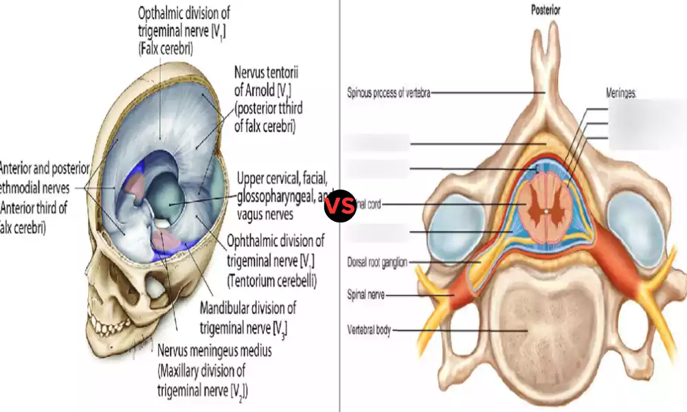
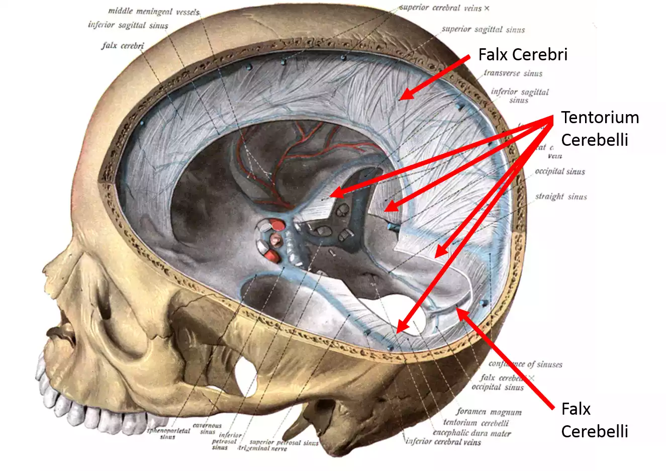
The cranial dura is split into two parts: the periosteal which is securely connected to the surface of the skull. The other layer is the meningeal, which is directly in contact with the surface of the brain. The dura mater also houses the dural sinuses. These help in the draining out of brain blood.
Definition of Spinal Dura
The spine dura mater is often just referred to as”the dura of the spine,” and comprises a tough fibrous membrane that encases and shields the spinal cord inside the spinal canal. It is among the three layers of meningeal tissue, alongside the arachnoid and pia mater.
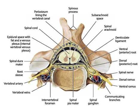
They form the protective cover over the nerve system of central control. The spinal dura comprises two distinct layers: an outer that is connected to the spine’s periosteum and the inside layer, that is tightly connected to the surface of the spinal cord.
The spinal dura helps protect your spinal cord against damage caused by mechanical forces it also houses cerebrospinal liquid (CSF) in the subarachnoid cavity and helps to ensure the stability and security of the spine as it travels across the canal of vertebrae.
Importance of understanding the differences
Understanding the distinctions between the cranial dura and the spinal dura is vital for a variety of reasons in the fields of neurology, medicine, and neurosurgery.
Here are a few of the main reasons to know the distinctions between these two:
- Diagnostic and Treatment: Recognizing the distinct features of the spinal and cranial dura is crucial for accurate diagnosis in clinical practice and treatment plans. Disorders that affect these structures, like dural herniations or tears, require specific treatment strategies.
- Surgery Procedures: Surgery specialists operating within the cranial and spinal regions require a clear understanding of the differences between dura mater to carry out procedures in a safe and efficient manner whether it’s cranial surgery like craniotomies or spinal surgeries such as laminectomies.
- Pathological conditions: Distinctive pathological conditions may affect the cranial or spinal dura. Knowing these differences aids in the identification and treatment of diseases such as Dural tumors, infection, or hemorrhagic hematomas.
- Cerebrospinal Fluid (CSF) Dynamics: Understanding the dural distinctions is essential to comprehend CSF dynamics. The cranial dura is home to structures similar to the dural sinuses that are involved with CSF absorption, and the spinal dura is a part as well in the CSF circulatory system and regulates pressure.
- Neurological Evaluation: Healthcare professionals and neurologists need to recognize the involvement of the spinal dura and the cranial nerve when evaluating symptoms of neurological conditions.
- Radiological Interpretation: Radiologists who interpret imaging studies, like MRIs as well as CT scans, must know these differences so that they can precisely identify any abnormalities that may be related to the dura and other structures around it.
- The research process: Understanding these differences is essential for researchers working in neuroanatomy, neuroscience, and neurophysiology. It is the basis to study different parts of the nervous system’s central.
- Patient education: Educating patients about the difference between cranial as well as spinal dura is a great way of increasing their understanding of surgical and medical issues which can improve their decision-making abilities and adherence to treatment plans.
A thorough knowledge of the difference between spinal and cranial dura is vital in the field of clinical practices, surgery and diagnostics, research, and patient care in the fields of neurology as well as neurosurgery. It helps improve the outcomes of patients as well as improved surgical procedures as well as advancements in neuroscience science and medical science.
Comparison Table of Cranial Dura and Spinal Dura
Here’s a comparison table highlighting the key differences between Cranial Dura and Spinal Dura:
| Aspect | Cranial Dura | Spinal Dura |
|---|---|---|
| Location | Surrounds the brain within the cranial cavity. | Envelopes the spinal cord within the vertebral canal. |
| Layers | Comprises two layers: the Periosteal Layer (outer) and the Meningeal Layer (inner). | Comprises two layers: The outer Layer (continuous with vertebral periosteum) and the Inner Layer (adjacent to the spinal cord). |
| Direct Contact with Nervous System | The meningeal layer of the cranial dura is in direct contact with the brain’s surface. | The inner layer of the spinal dura is closely associated with the spinal cord’s surface. |
| Functions | Protection of the brain from external trauma. – Support and stabilization of the brain within the cranial cavity. – Houses dural sinuses for venous blood drainage. | Protection of the spinal cord from mechanical damage. – Containment of cerebrospinal fluid (CSF) within the subarachnoid space. – Contribution to spinal cord stability within the vertebral canal. |
| Cerebrospinal Fluid (CSF) | The cranial dura does not directly enclose CSF but contains dural sinuses involved in CSF absorption. | The spinal dura surrounds the subarachnoid space, where CSF circulates and exerts pressure on the spinal cord. |
| Surgical Procedures | Relevant in cranial surgeries like craniotomies and procedures involving the brain. | Relevant in spinal surgeries like laminectomies and procedures involving the spinal cord and nerves. |
| Clinical Conditions | Dural tears – Dural sinuses – Cranial dura pathologies | Dural herniations – Spinal meningoceles – Spinal dura pathologies |
| Radiological Importance | Important for interpreting cranial imaging studies, such as CT scans and MRIs. | Important for interpreting spinal imaging studies, such as spinal MRIs and CT scans. |
Understanding these differences is essential for healthcare professionals involved in the diagnosis, treatment, and management of conditions related to the central nervous system, whether they pertain to the brain (cranial dura) or the spinal cord (spinal dura).
What is the dura mater attached to?
The dura mater is part of the meninges that surround the brain and spinal cord, and connects to a variety of structures, based on the location of its attachment:
- Cranial Dura Mater (Cranial Dura):
- The periosteal lining of the cranial dura mater is connected to the inside of the skull. It is firmly attached to the bone of the vault of the cranial.
- The meningeal layer of the dura of the cranial area is directly in contact with the brain’s surface. It is separated from the periosteal by the space that is known as the “dural border cell layer.”
- Spinal Dura Mater (Spinal Dura):
- The dura mater, the outer skin layer on the spinal cord runs with the periosteum in the vertebral column. It is securely fixed to the interior surface that forms the spinal canal giving stability and protection to the cord.
- The layer that is inside the dura mater of spinal nerves is tightly linked to the surface of the spinal cord and covers the subarachnoid space that is filled with cerebrospinal liquid (CSF).
The dura mater’s connection to bones (skull and the vertebral column) is crucial in ensuring stability and protection for the nervous system’s central nerve. The dura mater functions as a barrier to protect the brain which prevents the spinal cord and brain from coming into contact with the hard bone structures in the skull as well as the vertebral canal.
What is absent in the dura mater of the spinal cord?
The spinal cord’s dura mater does not have an exact structure located in the cranial mater. Within the dura mater of the cranial region, it is home to venous structures, referred to by the name of “dural sinuses,” which are responsible for the draining of venous blood away from the brain. The dural sinuses including the superior sagittal and transverse sinuses are created through the division between the meningeal and periosteal layers of the cranial dura.
The spine’s dura mater doesn’t contain dural sinuses that are similar to the ones in the spinal cord. Although the spinal dura is connected to the cranial dura doesn’t have a special drainage system for venous blood. The primary purpose of the spine is to protect the spine from mechanical injury and to seal the subarachnoid cavity, in which the cerebrospinal fluid (CSF) flows around the spine.
What is missing from the dura mater that surrounds the spinal cord is the dural sinuses which are typical of the dura mater of the cranial region.
How does the dura mater provide protection?
The dura mater offers protection for the nervous system of central origin, which includes the spinal cord and brain via a number of mechanisms:
- Mechanical Security: The dura mater is a strong and fibrous membrane that covers the brain in the cranial cavity, as well as the spinal cord in the spinal canal. Its tough, supple nature creates a physical barrier that protects these essential nervous system structures from external pressures and trauma. It prevents injuries that may result from compression or collision.
- Encapsulation: Dura Mater covers the spinal cord and brain effectively, segregating them from other structures and tissues. This separation helps to maintain the integrity and functionality of the nervous system central by blocking interference from nearby structures.
- Cerebrospinal Fluid (CSF) Containment: When the spine is enclosed by the dura it surrounds the subarachnoid area that is filled with cerebrospinal liquid (CSF). CSF acts as a cushion that provides the buoyancy needed for shock and absorption for the spine, thus reducing the chance of injury caused by abrupt movement or sudden jolts.
- Protection against Infections: Dura mater creates an effective barrier to stop the spread of pathogens such as viruses or bacteria, from infiltrating the spinal cord or brain. It is vital to ensure sterility essential to maintain proper nerve system function.
- Venous Drainage (Cranial Dura): In the cranial dura mater, special dural sinuses, which are venous structures, aid in the removal of blood venous out of the brain. The venous drainage system plays an important role in maintaining the proper intracranial pressure as well as cerebral blood flow and contributes indirectly to the protection of the brain by controlling these crucial parameters.
The dura mater offers multiple protections to the nervous system central functioning as a physical barrier isolating neural tissue, which contains CSF and in the cranial area, helping to regulate the pressure inside the cranium and the drainage of venous blood. These protective functions are vital to maintain the health and functioning of the spinal cord and brain.
Similarities Between Cranial Dura and Spinal Dura
There are some notable distinctions between the cranial dura (dura mater in the brain) and the spinal dura (dura mater of the spinal cord) however, they share certain important features:
- Fibrous Composition: Both the cranial and the spinal dura are comprised of fibrous, tough connective tissue. The fibrous nature of the tissue offers strength and durability to these membranes of protection.
- Meningeal Layers: The spinal dura and cranial dura are made up of two layers:
- The Periosteal layer, the outermost layer is continuous with the periosteum of the skull (in the cranial dura) or the vertebral column (in the spinal dura).
- Meningeal tissue which is the inner layer, is directly in connection with neural tissues. In both instances it creates an insulating barrier around the spinal cord or brain.
- Security: The primary function of both cranial as well as spinal duras is to shield the neural structures beneath. They act as physical barriers, which protect your cerebellum (in the dura cranial) as well as the spinal cord (in the spinal dura) from mechanical force external to them like injury or trauma.
- Stability and Support: The dura mater structure and both aid in the stability and support of the neural tissue that they surround. The cranial dura assists in maintaining the location of the brain in the cranial cavity while the spinal dura offers stability for the spinal cord in the spinal canal.
- Attachment to skull structures: The cranial as well as spine dura maters are joined to bone structures. The cranial dura is attached to the outer part of the skull and the spinal dura is attached to the part of the vertebral canal. These attachments ensure the structural integrity of the central nervous system.
- Protection from Infection: The dura mater acts as a protective barrier to protect the neural tissue from infection by preventing pathogens from getting into the spinal cord or brain.
While there are some similarities however, it is important to remember that the spinal dura and the cranial also possess distinct functions and characteristics that have been described in prior responses. Knowing their similarities and differences is vital to gaining an in-depth understanding of their roles in protecting the central nervous system.
Conclusion
Both the cranial and the spinal dura have a lot in common with their fibrous structures and meningeal layer, as well as their protecting functions as well as support, attaching to bone structures, and the role they play in preventing infections.
They have distinct functional and anatomical differences which are crucial to comprehend when it comes to surgery, clinical practice as well as research into neurological disorders since they play vital roles in defending and sustaining the brain and the spinal cord.