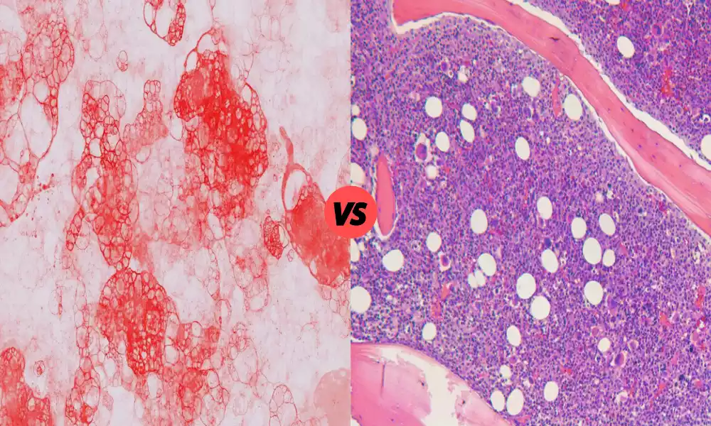
Osteosarcoma is a rare, extremely aggressive type of bone cancer that covers different subtypes, such as parosteal and periosteal osteosarcoma. The distinction between these subtypes is essential for a customized diagnosis and treatment.
The main Periosteal and Parosteal Osteosarcoma differences and similarities of parosteal and periosteal osteosarcomas, clarifying their clinical manifestation Histological characteristics, diagnostic issues, and treatments, all the while highlighting the importance of early and precise diagnosis when it comes to dealing with these difficult malignancies.
What is Periosteal Osteosarcoma?
Periosteal osteosarcoma can be described as a unique kind of bone cancer that develops from the periosteum, which is the outermost layer of bone, also known as periosteum. The periosteum is a fine but hard membrane that protects bones and plays a part in the healing and growth of bones. Periosteal osteosarcoma is a specific type of cancer that develops cells in or near the periosteum.
The subtype of osteosarcoma described above is distinguished by its position on the outside of bones and is most often found on the longer bones in legs and arms including the femur (thigh bone) or the tibia (shinbone). Periosteal osteosarcoma can be considered less dangerous than other types of osteosarcoma. However, there is still a need for immediate evaluation and therapy due to its ability to spread and cause major tissue and bone damage.

What is Parosteal Osteosarcoma?
Parosteal osteosarcoma is a rare type of bone cancer that arises from the bone’s outer tissue, also known as the periosteum. It is located close to the surface of the bone. In contrast to other kinds of osteosarcoma, which originate inside the bone, parosteal osteosarcoma is found on the outside of the bone.
The subtype of osteosarcoma described above is generally characterized by its slow growth rate and moderately low aggressiveness in comparison to other types of osteosarcoma. It is most often found on the long bones of extremities like the femur (thigh bone) or the tibia (shinbone). Parosteal osteosarcoma usually manifests as a bony, well-defined lump or mass on the surface of the bone.
While parosteal osteosarcoma is generally believed to have a higher prognosis than other kinds of osteosarcoma, it requires timely diagnosis and treatment because it is able to spread, invade surrounding tissues, and even result in complications if not treated.
Importance of distinguishing between periosteal and parosteal osteosarcoma
The distinction between parosteal and periosteal osteosarcoma is vital for a variety of crucial reasons:
- Plan of Treatment: The decision to choose treatment for osteosarcoma depends on the subtype of the disease. Parosteal and periosteal osteosarcomas can react differently to various treatment options, like chemotherapy, surgery, or radiation therapy. An accurate diagnosis can ensure that the patients get effective and appropriate treatment for their particular situation, which could result in better outcomes and reduce unwanted adverse negative effects.
- Prognosis: The prognosis as well as long-term results for patients with osteosarcoma may differ among types. Knowing which type one’s patient is able to provide important information about the course expected of disease as well as the probability of recurrence, as well as the possibility of metastasis (spread across other areas in your body). This information is vital for health professionals in making an informed decision in setting achievable expectations.
- Surgical approach: The surgical resection procedure is the most common treatment for both parosteal as well as periosteal osteosarcoma. But, the procedure may differ depending on the type of cancer and the site of the tumor. The distinction between these two types assists surgeons in planning the best surgical procedure to complete the removal of the tumor while keeping as much bone health and function as they can.
- Research and Clinical Trials: Different subtypes of osteosarcoma can respond differently to the latest therapies and treatments. Research and clinical trials usually focus on particular types of cancer. A precise classification of periosteal and parosteal osteosarcomas ensures that patients are able to participate in relevant clinical trials. It can also help in the advancement of knowledge as well as the creation of new therapies.
- Patient Education and Assistance: Understanding the specific osteosarcoma subtype can provide the patients as well as their family members with information about the condition, its features, and treatment options. This knowledge will result in more informed choices and better compliance with treatments, which can improve the overall well-being of patients.
Discerning between osteosarcomas that are parosteal and periosteal is vital for tailoring treatments, predicting outcomes improving surgical techniques assisting research efforts, and giving patients and their families the support and information they require throughout their journey with cancer.
Comparison Table of Periosteal and Parosteal Osteosarcoma
Below is a comparison table highlighting the key differences between periosteal and parosteal osteosarcoma:
| Characteristic | Periosteal Osteosarcoma | Parosteal Osteosarcoma |
|---|---|---|
| Origin | Develops on the periosteum, the outer layer of the bone. | Develops adjacent to the periosteum on the bone’s surface. |
| Location | Typically occurs in the long bones of the extremities, such as the femur or tibia. | Also typically found in the long bones, often in the posterior aspect of the femur. |
| Growth Pattern | Tends to grow away from the bone, with a “mushroom-like” appearance. | Grows on the external surface of the bone, often forming a distinct, well-defined mass. |
| Aggressiveness | Generally considered less aggressive than other osteosarcoma subtypes. | Relatively slow-growing and less aggressive compared to other subtypes. |
| Histological Features | Typically displays characteristic features under the microscope. | Histologically distinct, with well-differentiated features. |
| Clinical Presentation | May present with pain, swelling, and limited joint movement. | Often presents as a painless, firm lump or mass near a bone. |
| Metastasis Risk | Has a lower risk of metastasis (spreading to other parts of the body) compared to other subtypes. | Metastasis is less common but can occur if left untreated. |
| Treatment Approach | Primary treatment involves surgical resection. Chemotherapy may be used depending on the extent of the disease. | Surgical removal is the main treatment, with chemotherapy being less frequently used. |
| Prognosis | Generally associated with a better prognosis compared to other osteosarcoma subtypes. | Also associated with a favorable prognosis, but early and accurate treatment is still critical. |
It’s important to note that while periosteal and parosteal osteosarcomas have their unique characteristics, they are both considered rare forms of osteosarcoma, and individual cases can vary in terms of presentation and treatment response. Patients diagnosed with either subtype should receive personalized care and treatment plans tailored to their specific condition.
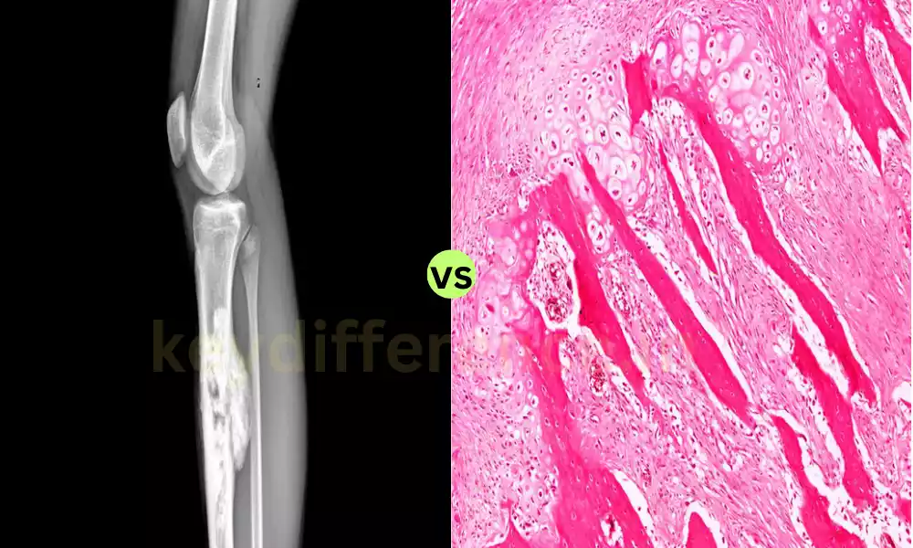
MRI and CT scan characteristics
MRI as well as CT scan characteristics may aid in identifying the difference between periosteal and parosteal osteosarcomas since imaging results can provide useful information about the location, size, and features of the cancers.
Here are a few typical MRI as well as CT scan characteristics that apply to both types:
Periosteal Osteosarcoma:
MRI Characteristics:
- Tumor Localization: Periosteal osteosarcoma typically is seen as a lump that lies on the outside of the bone affected.
- Signal Intensity: On MRI the periosteal osteosarcoma usually displays a variety of signal intensity in T1-weighted and T2-weighted MRI images.
- Soft Tissue Mass: The tumor can expand into the soft tissues surrounding it.
- Enhancement: After contrast administration (contrast-enhanced MRI), there is typically significant improvement in the cancer.
CT Scan Characteristics:
- Mineralization: Periosteal osteosarcoma can exhibit zones that are mineralized (calcification) inside the cancer. These could be seen as hyperdense areas when viewed on CT scans.
- Bony Changes: The tumor can trigger changes to the bone adjacent like thickness of the cortical or erosions.
Parosteal Osteosarcoma:
MRI Characteristics:
- Well-Demarcated Mass: Parosteal osteosarcoma typically appears as a well-demarcated, lobulated, and well-demarcated lump near the bone.
- Lower to Intermediate Signal: On MRI the signal strength of parosteal osteosarcomas is usually low to intermediate in T1-weighted images. It is also heterogeneous when T2-weighted images.
- Minimum Soft Tissue Involvement: It is a condition that has very little involvement of the soft tissues surrounding it.
- Enhancement: As with periosteal osteosarcoma Parosteal osteosarcoma could be enhanced after the administration of contrast.
CT Scan Characteristics:
- Exophytic Growth: Parosteal osteosarcoma typically displays an exophytic pattern of growth, that extends outwards from bone.
- Mineralization: Similar to osteosarcoma periosteal, regions of calcification or mineralization may be detected in CT scans.
- Changes in adjacent bone: The tumor can result in changes to the bone adjacent to it including cortical thickness, however, without significant bone loss.
It’s crucial to keep in mind that, while these characteristics of imaging can be useful in determining the diagnosis, however, the absolute diagnosis for osteosarcoma can be generally confirmed by an amalgamation of histological analysis, imaging of a biopsy sample, and a clinical assessment. the individual cases could differ, and the evaluation of the results of an imaging scan is best handled by experienced radiologists and oncologists.
Prognosis and treatment approaches
Treatment and prognosis for periosteal or parosteal osteosarcoma may differ, however, both types tend to be more successful when compared with different, less aggressive types of osteosarcoma. Here’s a summary of the prognosis and treatment options for each type:
Prognosis:
- Periosteal Osteosarcoma:
- The prognosis for periosteal osteosarcoma generally is favorable and has a better long-term survival rate when compared to other osteosarcoma types.
- The 5-year survival rate of the periosteal osteosarcoma may range from 70% to 90 percent.
- The early diagnosis and the appropriate treatment can significantly improve a favorable prognosis.
- The chance that metastasis (spread to distant organs) is less in osteosarcoma with periosteal insertions in comparison to other subtypes.
- Parosteal Osteosarcoma:
- Parosteal osteosarcoma can also have a positive prognosis.
- 5-year survival rates of parosteal osteosarcoma tend to be high, sometimes at or above 90%…
- Like periosteal osteosarcomas and periosteal cancer, diagnosis, and treatment play an important role in achieving a positive result.
- While metastasis is not as common in parosteal osteosarcomas. However, however, it could occur when the tumor is untreated or not completely removed.
Treatment Approaches:
- Periosteal Osteosarcoma:
- Surgery Resection: The main procedure for treating periosteal osteosarcoma consists of surgery to remove the cancer while keeping as much healthy bone as is feasible. Limb-sparing surgery can be performed in many cases.
- Chemotherapy: Depending upon the size and nature of the cancer, periosteal osteosarcoma is treated with chemotherapy before and following surgery.
- Radiation Therapy: The use of radiation therapy is not often utilized as a primary treatment, however, it could be considered in some instances.
- Parosteal Osteosarcoma:
- Surgery: Surgery is the primary form of therapy for patients with parosteal osteosarcoma with the intention of removing the tumor completely while maintaining joints and bones.
- Chemotherapy: In contrast to other types of osteosarcomas, parosteal osteosarcoma is more resistant to chemotherapy and its usage is limited. It is typically reserved for instances in which surgery alone is inadequate.
- Radiation Therapy: Treatment with radiation is usually not considered a primary treatment, but it can be considered in some circumstances.
There is a choice of treatment and the method of treatment will depend on aspects like the size, the location, its extent as well as the patient’s health and needs. Multidisciplinary teams consisting of orthopedists, medical oncologists, and radiologists work together to create specific treatment plans designed to get the most effective result while maintaining limb functioning and living quality.
Regular follow-up and monitoring are essential to look out for the possibility of metastasis or recurrence and adjustments to treatment plans can be required to achieve the most effective long-term outcomes.
Importance of early detection
The early detection of both parosteal and parosteal osteosarcomas is of crucial importance due to several important reasons:
- Better Treatment Results: Finding these subtypes of osteosarcoma at the earliest stage usually leads to better treatment outcomes. Lesser tumors are typically simpler to remove surgically and are less likely to be able to spread to other parts of the human body. This may increase the chances of complete removal of the tumor and a greater chance of long-term longevity.
- Limb-Sparing Surgery: Early detection can lead to a limb-sparing procedure, in which the bone cancerous segment can be removed while maintaining the function of the limb affected. This is crucial to ensure the patient’s well-being, mobility, and autonomy.
- Low Treatment Intensity: When the tumor is identified earlier treatment options that are less aggressive could be contemplated. This may result in fewer adverse effects and a higher general quality of life for the patient.
- Prevention of Metastasis: Osteosarcomas when left untreated can spread into other organs making them more difficult to treat. Early detection can stop or minimize the chance of metastasis by treating the tumor prior to it having an opportunity to spread.
- Improved Survival Rates: Osteosarcoma rate of survival is considerably more favorable when the cancer is discovered in its earliest stages. The early intervention and proper treatment can significantly improve the chances of a patient’s long-term survival.
- Reduced Risk of Complications: Osteosarcoma in the advanced stage can cause complications, such as the development of pathological fractures and extreme discomfort. The early detection of and treatment could aid in reducing these complications as well as the suffering that comes with them.
- Quality of Life for Family and Patient: Early detection provides patients and their families with a sense of control as well as the chance to participate actively in the treatment decision-making process. It also eases the psychological and emotional stress that comes with the delay in diagnosis.
- Researchers as well as Clinical Trials: Patients diagnosed with osteosarcoma that are in its infancy may have the chance to participate in clinical trials, and help in the creation of new treatments and options for these uncommon cancers.
In order to facilitate the early detection of cancer to prevent early detection, healthcare professionals and patients should be on the lookout for the signs or symptoms that indicate osteosarcoma for example, chronic swelling, pain in the bone, or undiagnosed fractures.
A timely medical examination as well as imaging tests (e.g. scans, X-rays MRI, CT scans) are a great aid to the detection of these cancers.
The increased awareness among healthcare professionals, especially radiologists and orthopedic surgeons could result in more timely and accurate diagnoses, thereby improving the chances of those who are affected by parosteal or periosteal osteosarcoma.
Ongoing research and advancements in osteosarcoma management
Continuous research and advances in the management of osteosarcoma are essential in improving the treatment, diagnosis, and results of patients who are affected by this uncommon and highly aggressive type of bone cancer.
Here are a few areas of ongoing research as well as recent advances in osteosarcoma treatment:
- Precision Medicine and Targeted Therapies: Researchers are exploring the possibility of using targeted therapies that target molecular or genetic changes that occur within osteosarcoma tumors. This strategy aims at tailoring treatment according to the specific features of each individual patient’s tumor which could increase the effectiveness of treatment and minimize the risk of side consequences.
- Therapy for Immunity: Therapies that target the immune system, including immune checkpoint inhibitors, as well as the chimeric antigen receptor (CAR) treatment with T-cells are being studied to see if they could make use of the body’s immune system to destroy and target osteosarcoma-related cells.
- Genomic Profiling: Recent advances in genetics have enabled more thorough analysis of the genome for osteosarcoma tumors. Understanding the genetic mutations that cause modifications within these tumors may result in more targeted treatment as well as the creation of new treatments.
- Improved Radiation Therapy: Techniques for delivering radiation therapy have improved to allow for more specific and precise radiation to protect healthy tissues while efficiently treating tumors. This is especially beneficial in cases of osteosarcoma in which radiation therapy is recommended.
- Research and Development in Drugs: Ongoing research efforts concentrate on the design of innovative treatments and drugs specifically formulated to fight osteosarcoma. This includes novel chemotherapeutic drugs and combinations of therapies that could boost the effectiveness of treatment.
- Biomarker Identification: The identification of biomarkers that are associated with osteosarcoma may assist in diagnosing the disease early, and predicting prognosis and treatment choice. Researchers are currently working on finding reliable biomarkers for this type of cancer.
- Clinical trials: A variety of clinical trials are currently underway to test the safety and effectiveness of innovative treatment options for osteosarcoma. These trials typically include research therapies, immunotherapies, and cutting-edge surgical methods.
- Minimally Invasive Surgery: Modern advances in surgical methods are examining the possibility of using minimally invasive techniques for the removal of tumors which reduces the effect on healthy tissue surrounding it and possibly improves the speed of recovery.
- Support for Patients and Advocacy: Patient advocacy groups and other organizations are working to educate the public about osteosarcoma provide assistance to those affected as well as their families, and encourage participation in clinical studies.
- Collaboration Research Networks: Collaborative efforts of medical institutions, researchers, and international networks help connect resources, share data, and speed up progress in osteosarcoma research.
The ongoing research efforts and developments in osteosarcoma management are promising for improving the treatment and diagnosis of this difficult cancer. As our knowledge of the disease advances and improves, we are hopeful that these efforts result in better outcomes, higher rates of survival, and better health for patients who are diagnosed with osteosarcoma.
Similarities Between Periosteal and Parosteal Osteosarcoma
Periosteal and parosteal osteosarcomas are two types of osteosarcoma with distinctive characteristics, they have a few similarities:
- The source of HTML0 is the Periosteum: Both periosteal osteosarcoma and parosteal osteosarcoma cancers arise from or are a part of the periosteum the thin layer that covers the outside of bones. This is the most distinctive characteristic of both types.
- Placement in Long Bones: Both osteosarcomas periosteal and parosteal are likely to be found in bone that is long in extremities like the femur (thigh bone) or the tibia (shinbone). Although the exact position on the surface of the bone may be different, however, their preference for long bones shares a commonality.
- Metastatic Low to Intermediate Potential: In comparison with other, more aggressive types of osteosarcoma, both parosteal and periosteal osteosarcomas have the lowest risk for metastatic spread (spreading into distant organs). However, metastasis may be a possibility if the tumor is not treated or when the tumor isn’t completely eliminated.
- Image Characteristics: Based on imaging and radiographic characteristics, both periosteal as well as parosteal osteosarcomas might show regions of mineralization or calcification inside the tumor. These areas are visible in X-rays or CT scans.
- The importance of early detection: Early detection and diagnosis are crucial for both parosteal and periosteal osteosarcoma. The detection of these subtypes early stage could result in better results in treatment and a greater probability of success in surgical removal.
- Treatment Modalities: The most common treatment method for both types is surgical resection with the goal of removing the tumor and preserving as much bone health and function as much as is feasible. Chemotherapy may be considered in certain instances, but it is not used as often as other subtypes of osteosarcoma.
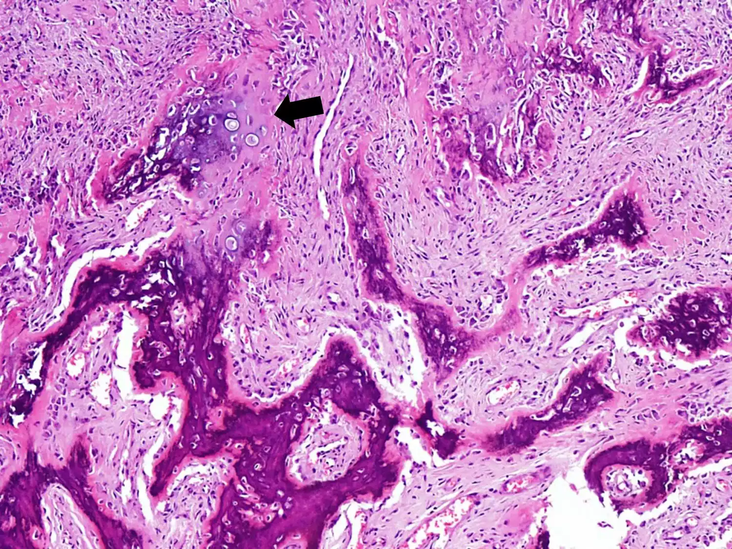
- The Histological Basis: Both periosteal and parosteal osteosarcomas can be classified as osteosarcoma subtypes, with the same histological ancestry in bone-forming cells (osteoblasts as well as their precursors).
Despite the similarities, it is important to be aware that periosteal as well as parosteal osteosarcomas have distinct distinctions, such as their development patterns, histological features, and clinical manifestations.
An accurate diagnosis and distinction between these types is essential to tailor treatment plans according to the specific patient’s needs and achieve the most effective results.
Reference Books
Certainly! Here are some reference books on osteosarcoma and related topics:
- “Osteosarcoma: Diagnosis and Treatment” by Robert J. Grimer and Stephen R. Cannon
- This book provides a comprehensive overview of osteosarcoma, covering topics such as diagnosis, treatment options, surgical techniques, and outcomes.
- “Bone and Soft Tissue Tumors: A Multidisciplinary Review with Case Presentations” by George W. J. Keeney and Santiago A. Lozano-Calderon
- This text offers a broader perspective on bone and soft tissue tumors, including osteosarcoma, with case presentations and insights from a multidisciplinary team of experts.
- “Bone Cancer: Primary Bone Cancers and Bone Metastases” edited by Dominique Heymann and Franz E. Weber
- This book explores various aspects of bone cancer, including osteosarcoma, with a focus on both primary bone cancers and bone metastases. It covers molecular biology, pathology, diagnosis, and treatment strategies.
- “Orthopedic Oncology: Diagnosis and Treatment” edited by Ernest U. Conrad, Frank J. Frassica, and Donald W. Love
- This comprehensive reference text covers a wide range of topics related to orthopedic oncology, including osteosarcoma, with a focus on diagnosis, treatment options, and surgical techniques.
- “Pediatric Bone and Soft Tissue Sarcomas” edited by Alberto S. Pappo and Cindy L. Schwartz
- This book focuses specifically on bone and soft tissue sarcomas in pediatric patients, including osteosarcoma. It discusses pediatric-specific considerations in diagnosis and treatment.
Conclusion
osteosarcoma, and its subtypes like periosteal or parosteal osteosarcoma, pose distinct challenges for the diagnosis and treatment. The ability to differentiate between these types and early detection is crucial to improve the patient’s outcomes.
Innovations in the field of research and treatment strategies which include precision medicine as well as immunotherapy, have the potential of improving the prognosis and quality of life for patients suffering from osteosarcoma.
Although ongoing efforts are being made to improve our understanding and treatment of this rare bone cancer collaboration across disciplines and patient care is essential to the path toward better outcomes and eventually finding an eventual cure.