
The eye of the human being is an extremely intricate organ that has numerous different layers, and various structures that work to enhance vision. Choroid and Sclera of the most important elements in the structure of an eye are the sclera and the choroid. Both play a vital role in maintaining the health of your eyes however, they are distinct in terms of structure as well as function and the location in the eye.
Understanding the differences between them is crucial to understanding the complex workings of the eye as well as the treatment and diagnosis of ocular diseases. We’ll examine the differences between the sclera and the choroid in order to better understand their unique role in maintaining vision as well as general eye functioning.
What is Choroid?
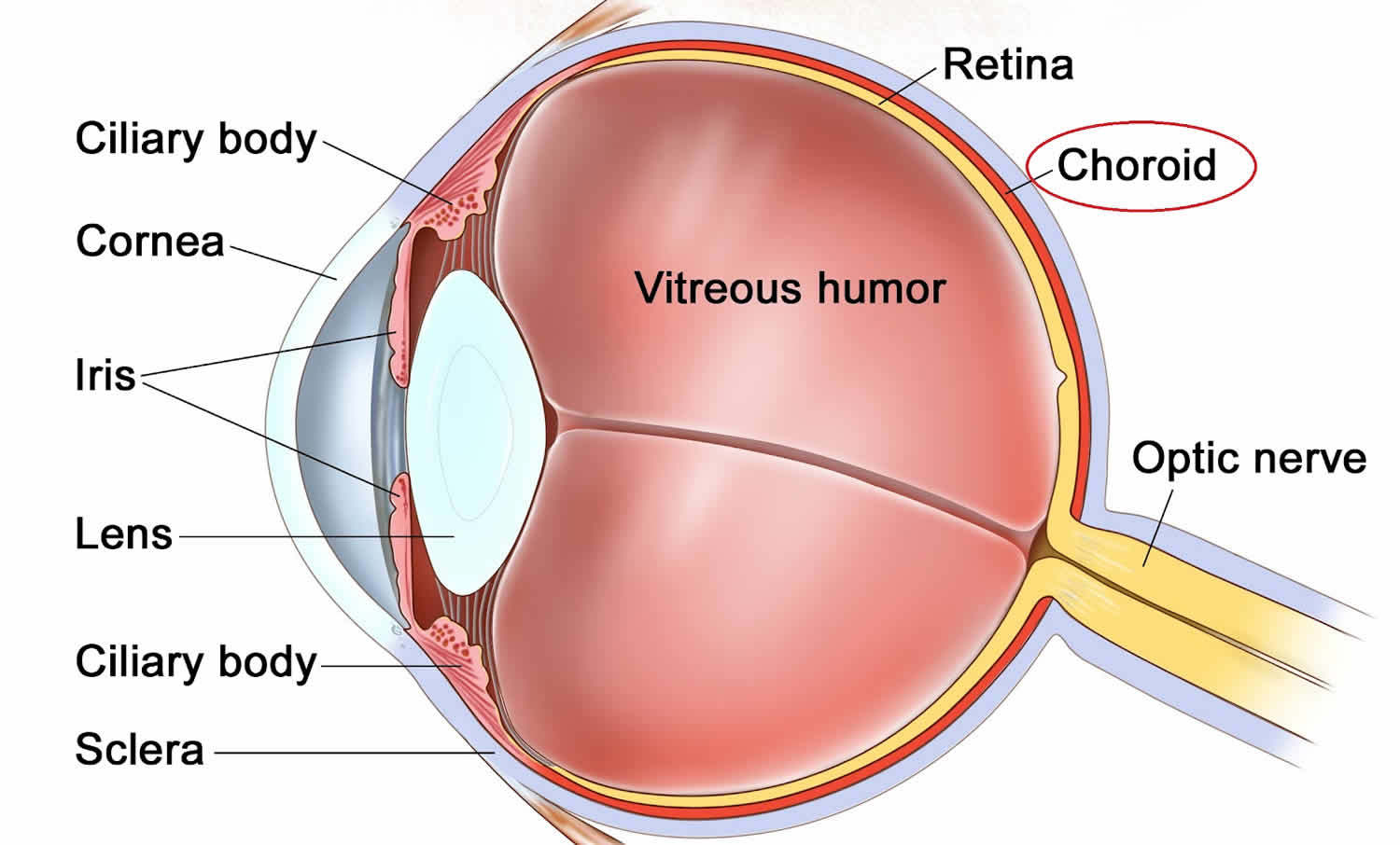
The choroid is a densely vascular tissue within the eye, primarily in the middle of the eyeball. It lies within the eyeball between the retina (the inner layers) as well as the sclera (the outermost layer). It consists of pigment cells, blood vessels, and nerves.
The choroid plays an essential function in providing oxygen and nutrients to the retina, which are vital to maintaining the health and functioning of the photoreceptors responsible for vision.
It helps maintain the eye’s temperature and absorbs light to reduce reflections and glare in the eye, thereby enhancing seeing clarity.
What is Sclera?
The sclera often called”the “white of the eye,” is the tough, opaque, and fibrous outer part of the eyeball. It creates the transparent, protective outer layer of the eye. It also protects the delicate structures inside like the corneas, the iris, and the lens.
The main purpose of the sclera is to give structural support and keep the shape of the eye, as well as protect internal structures from damage caused by external forces.
It serves as an anchor to the muscles that control eye movement. Its strength and durability protect the delicate structures inside the eye which allows them to function at their best.

Comparison Table of Choroid and Sclera
Here’s a comparison table highlighting the key differences between the choroid and sclera:
| Characteristic | Choroid | Sclera |
|---|---|---|
| Location in the Eye | Located between the retina and sclera | The outermost layer of the eyeball, forming the white part of the eye |
| Composition | Highly vascular with blood vessels, pigment cells, and nerves | Dense connective tissue, primarily collagen fibers |
| Function | – Nutrient supply to the retina – Thermoregulation – Absorption of excess light | – Protection of inner eye structures – Maintenance of eye shape and integrity – Attachment point for eye muscles |
| Appearance | The dark, vascular layer | White, tough, and opaque |
| Role in Vision | Indirect role by supporting the retina | No direct role in vision but provides structural support |
| Clinical Significance | Choroidal diseases and conditions include choroiditis and choroidal neovascularization | Scleral diseases and conditions include scleritis and scleral thinning |
This table summarizes the fundamental distinctions between the choroid and sclera, emphasizing their unique compositions, functions, appearances, and clinical relevance in eye health.
Importance of understanding the differences between choroid and sclera
Knowing the distinctions between the sclera and the choroid is crucial for a variety of reasons:
- Diagnostic and Treatment: Ophthalmologists and other healthcare professionals depend on their understanding of the anatomy of the eyes to diagnose and treat a variety of eye diseases and conditions. Differentiating between the choroid and sclera assists in identifying the root of any potential issues and is vital for accurate diagnosis and effective treatment.
- Eye Health Management: Knowing about the eye’s structures allows people to understand the intricate nature of the eye as well as the significance of ensuring its health. This knowledge could result in better eye health procedures and the early detection of eye problems.
- Surgery Procedures: Surgeons performing eye operations must be proficient in the eye’s anatomy to perform procedures with safety and efficiency. The understanding of the choroid as well as the sclera is vital in any surgery that involves the posterior portion of the eye.
- Vision and Comfort: A basic understanding of how the choroid scavenges excessive light and the way that the sclera supports structurally will help us understand the way our eyes work to give comfortable and clear vision. This knowledge is valuable for people who are looking for ways to shield their eyes from the glare while maintaining excellent vision.
- Information and Education: The process of educating the population about the anatomy of the eyes such as the choroid and the sclera, will raise awareness of the importance of regularly scheduled examinations of the eyes in eye health, eye protection, and overall health of the eyes. This information can result in proactive measures to avoid eye problems.
- The research and development process: For researchers in the field of ophthalmology or related sciences of the eye, knowing the different aspects of the eye’s structures is crucial. It serves as a basis to explore the latest treatments, therapies, and techniques to treat conditions of the eye.
- Communication with healthcare providers: When patients understand the terms and basics of the eyes they are able to better communicate with their healthcare professionals ask questions with confidence and make informed decisions regarding their eye health.
Understanding the distinctions between the sclera and the choroid isn’t only important for health professionals, but also for those who want to keep their eyes healthy and make informed decisions regarding their eye health. It is a vital component in the diagnosis, treatment education, and overall eye health treatment.
Description of the choroid’s position within the eye
The Choroid is a layer of vascularity that is located inside the eye. It is located in between the retinas and sclera. It is located towards the rear of the eyes and is a part of the uvea that comprises the iris and ciliary body.
Here’s a more in-depth description of the choroid’s location in the eye:
- Posterior Choroid: The choroid is found in the posterior portion of the eye, and extends from the ciliary body as well as the ora serrata (a sharp boundary of the retina) in the front, and finally the head of the optic nerve (where the optic nerve leaves the eye) in the back.
- Adjacent to the Retina: It is located adjacent to the Retina The choroid is located directly below the retina and has an extremely thin Bruch’s membrane that separates the two. The choroid offers vital nutrition and support to the outermost layer of the retina, also known as”the photoreceptor” layer.
- Sits Behind the Sclera: The choroid also lies posterior to the sclera which is the tough, white exterior covering that covers the eyes. It is separated from the sclera through the connective tissue layer called the suprachoroid lamina.
- Vascular Layer: This layer is extremely vascular, which means it has a large system of blood vessels. The blood vessels perform a vital function in supplying nutrition and oxygen to the retina’s outer layers as well as ensuring the proper functioning of the retina.
- Light Regulation: In addition to its functions as a nutrient, the choroid also plays a role in regulating the amount of light entering the eye, by taking in excess light energy, and also decreasing light scatter within the eye.
The choroid’s strategic location within its eye is vital to maintaining the health and functionality of the retina. Additionally, it plays a role in the overall visual system by assisting retinal photoreceptors as well as aiding in controlling the levels of light in the eye.
Description of the sclera’s position within the eye
The sclera, or sclera, is a hard fibrous layer that is the outermost protective layer of the eye. It plays an important part in preserving the shape of the eye and safeguarding its sensitive internal organs.
Here’s a brief description of the sclera’s location in the eye:
- Sclera: This layer functions to act as an outer layer for the eyes. It encompasses the majority of the surface of your eyeball. It’s the portion of the eye that’s evident when you look at an individual’s eyes. It also creates the eye’s distinctive white appearance.
- Envelopes the Eye: The sclera covers the entire surface of the eyes, protecting the contents of the eye and providing the eye with sturdy, secure skin. The sclera extends out from the front, which creates the cornea transparent towards the back which surrounds the optic nerve when it leaves the eye.
- Continuous with Continuous with Cornea: In front of your eye, the sclera smoothly changes into the cornea which is the clear dome-shaped structure covering the pupil and the iris. The cornea is translucent which allows light to enter the eye, whereas the sclera remains opaque and creates structural strength.
- Attachment points: The eyelid offers attachment points for extraocular muscles that regulate how the eyes move. These muscles control the gaze of the eye in various directions.
- Protection and Maintenance of Shape: The primary purpose of the sclera is to shield the eyes’ delicate structures inside, including the choroid, retina, and vitreous humor, from damage or trauma external to them. In addition, the sclera plays an essential role in keeping the shape of the eye and also stopping it from collapsing.
- Communication of Forces: This sclera assists in transmitting and distributing the forces produced by the muscles of the eye during movements that ensure smooth and coordinated movement.
The sclera functions as the outermost part of your eye, offering protection, structural support, and connective points for the muscles of the eye. The white appearance of the sclera is an identifying characteristic of the eye.
It can be easily seen in its front, where it is the white visible surface and continues to the posterior side to shield and protect the entire eyeball.
Diagnostic methods for choroid disorders
Choroid-related disorders may cause damage to the vascular layer in the eye and could result in various eye problems and problems with vision. To determine if choroid issues are present eye doctors employ various diagnostic techniques and instruments.
Here are some of the most common ways to diagnose choroid problems:
- Comprehensive Eye Examination:
- An extensive eye exam conducted by an optometrist or ophthalmologist is usually the initial step in diagnosing choroid problems. It includes assessing pupillary and visual acuity and examining the internal and external components of your eye.
- Ophthalmoscopy:
- Ophthalmoscopes that are indirect and direct enable the eye doctor to look at the choroid, retina, as well as the optic nerve’s head. They utilize a specific instrument called an ophthalmoscope. It allows them to observe the choroid, and look for any signs of abnormalities, such as hemorrhages, tumors, or inflammation.
- Fluorescein Angiography:
- Fluorescein angiography involves injecting a fluorescein-colored dye (fluorescein) into an arm vein and then capturing images of the eyes using a specialized camera. The test can help visualize blood vessels within the retina and choroid. The abnormalities in blood flow and leakage are identified which aids in the identification of choroid diseases such as Choroidal Neovascularization (CNV) also known as central serous cerebroretinopathy (CSC).
- Indocyanine Green (ICG) Angiography:
- ICG angiography is comparable to fluorescein angiography, but it uses an alternative color (indocyanine green). It is especially useful for the examination of choroidal blood vessels that are deeper and can be used to determine if specific choroid issues are suspected.
- Optical Coherence Tomography (OCT):
- OCT is a non-invasive imaging method that offers cross-sectional images of the retina, choroid, and other eye structures. It can help in the diagnosis of problems with the choroid by assessing the thickness and quality of the choroid, as well as detecting abnormalities in tissue or fluids and tracking the changes in time.
- Ultrasound Imaging:
- In the event that the choroid is not able to be visible using conventional imaging techniques, Ultrasound images (B-scan or A-scan) could be used to evaluate the thickness and strength of the choroid.
- Visual Field Testing:
- Tests of visual field, including perimetry, evaluate the patient’s peripheral and central vision. These tests help to diagnose and track conditions such as choroiditis that can impact the sensitivity of visual fields.
- Blood Tests and Laboratory Investigations:
- In certain cases, the use of blood tests or laboratory tests is required to find the underlying conditions related to choroid issues like autoimmune diseases or infections.
- Electroretinography (ERG) and Electrooculography (EOG):
- These tests assess how the brain’s electrical circuits work. These tests can be used to assess the functioning of the choroid and the retina in the event of suspected choroid problems.
- Genetic Testing:
- In the event that the possibility of inherited choroid diseases is raised genetic testing could be used to pinpoint specific genetic changes that cause the condition.
The selection of the diagnosis method(s) is based on the particular symptoms and suspect diagnosis, in addition to the experience of the healthcare professional. A precise diagnosis is essential in the management and treatment of choroid-related disorders.
Diagnostic methods for Sclera disorders
The sclera can be a problem for the fibrous, tough exterior part of your eye. It could cause various eye problems and discomfort. To identify sclera-related disorders specialists in eye care use an array of clinical examinations as well as specialized tests and imaging methods.
Here are some typical screening methods for sclera-related disorders:
- Comprehensive Eye Examination:
- An eye examination that is thorough by an optometrist or ophthalmologist is often the first stage in identifying sclera-related disorders. This exam evaluates the pupils, and visual acuity and also evaluates the internal and external eye structures as well as their appearance.
- Slit-lamp Biomicroscopy:
- Slit-lamp biomicroscopy entails using special microscopes that have the shape of a slit to look at the sclera and the other structures of your eyes in greater detail. This method allows for an examination in a close-up of the tissue’s color, texture, and other irregularities.
- Ocular Surface Staining:
- Fluorescein, also known as rose eye drops could be used to color the surface of the eye, including the sclera. The dyes are able to highlight any irregularities like rough spots, dry patches, or scratches of the sclera. These could be a sign of a particular disorder.
- Ultrasonography (Ultrasound):
- Ultrasound imaging is a method to evaluate the structural strength and thickness of the sclera, when observing it directly is difficult. Ultrasound probes with high-frequency frequencies can offer precise images of the anterior segment, which includes the sclera.
- Biopsy:
- The need for scleral biopsies may be necessary in order to collect an appropriate sample of tissue for laboratory analysis. This is generally reserved for instances where inflammation or autoimmune disorders are suspected.
- Blood Tests and Laboratory Investigations:
- Tests for blood can be done to determine underlying disorders that can be linked to sclera including autoimmune diseases or infections.
- Imaging Modalities:
- Imaging techniques such as optical coherence imaging (OCT) or magnetic resonance imaging (MRI) can be utilized to evaluate the sclera as well as surrounding structures in more detail, especially in situations where more complex structures are affected, or when assessing the severity of the scleral disorder.
- Rheumatologic Evaluation:
- If an autoimmune disorder or rheumatologic one could be identified, a rheumatologic assessment could be required to evaluate the overall health of the patient and determine any other systemic issues that could be contributing to the sclera disorder.
- Dye Disappearance Test:
- The test consists of placing the color of a dye (e.g. fluorescein) on the sclera, and checking how quickly it goes away. It could be helpful in testing scleral permeability as well as the identification of issues that affect the integrity of scleral tissue.
- Corneal Topography:
- Although it is mostly used to evaluate corneal topography the corneal topography may also offer valuable information regarding the shape and curvature of the sclera.
The selection of the diagnosis method(s) is based on the symptoms that are specific to your diagnosis, and the experience of the healthcare professional. A precise diagnosis is crucial to determine the root causes of sclera-related disorders and determine the best treatment options.
Similarities Between Choroid and Sclera
The sclera and choroid are both vital components in the anatomy of the eyes and despite their distinct functions and structures, however, they share a few similarities.
Here are the most significant similarities between the choroid as well as the sclera:
- Both Are Part of the Uvea:
- The sclera and the choroid are both parts of the uvea. This forms the middle of your eye. The uvea comprises the eye’s iris, the ciliary body, the choroid, and the underside connective tissue layer which comprises the sclera.
- Structural Support:
- Both the sclera and choroid give structural support to the eye. The sclera, which is the outermost layer of the eye, helps keep the shape of the eye and shields the internal structures. The choroid which lies below the retina and sclera, is also a contributor to the structural integrity of the eye.
- Protection of Inner Eye Structures:
- Both tissues play a part in protecting the fragile eye’s internal structure, like the retina, the vitreous humor, and the optic nerve. The sclera serves as an outer layer of toughness and the choroid acts as the vascular layer that provides nourishment to the retina and helps maintain its health.
- Connective Tissue:
- The sclera and the choroid are made up of mainly connective tissue. The choroid is home to an extensive blood vessel network while the sclera comprises collagen fibers as well as fibroblasts, which provide it with its strong, fibrous appearance.
- Position in the Eye:
- The sclera and the choroid are located in the posterior segment of the eye. These extend out from the anterior of the eye all the way to the back with the sclera acting as the outermost layer, and the choroid beneath it.
- Contribution to Eye Function:
- The choroid plays an important role in feeding the retina and controlling the amount of light entering the eye, and the sclera provides structural protection, both tissues contribute to overall health and function in the eyes.
- White Appearance:
- The outer layer of sclera that is visible when you look at your eyes, gives it its distinctive white appearance. Choroids, while not visible, contribute to the overall appearance of the eye.
It’s crucial to understand that, despite the similarities, the sclera and the choroid both have their own distinct roles and structures inside the eye. Each has its own unique function to maintain eyesight and health.
Reference Books
Certainly! Here is a list of reference books across various genres and subjects.
These books cover a wide range of topics and can serve as valuable resources for learning and research:
1. Science and Nature:
- “Cosmos” by Carl Sagan
- “A Brief History of Time” by Stephen Hawking
- “The Selfish Gene” by Richard Dawkins
2. Literature and Fiction:
- “To Kill a Mockingbird” by Harper Lee
- “1984” by George Orwell
- “Pride and Prejudice” by Jane Austen
3. History:
- “A People’s History of the United States” by Howard Zinn
- “The Guns of August” by Barbara W. Tuchman
- “Sapiens: A Brief History of Humankind” by Yuval Noah Harari
4. Philosophy:
- “Meditations” by Marcus Aurelius
- “The Republic” by Plato
- “Critique of Pure Reason” by Immanuel Kant
Conclusion
Reference books are valuable resources that provide knowledge, insight, and inspiration on many different subjects. If you are looking to broaden your knowledge of literature, science and philosophy, history, or any other subject the reference books offer many sources of information.
They also can serve as guides to continuous research and study. With the appropriate reference books available you are able to embark on intellectual adventures, gain new perspectives, and expand your knowledge about the globe.